

Description
The latest technology for perfect images with high diagnostic value.
Thanks to the introduction of the most recent technology and the newest features in digital radiology, X-VIEW 2D PAN requires a minimum exposure time to capture clear and contrasted images.
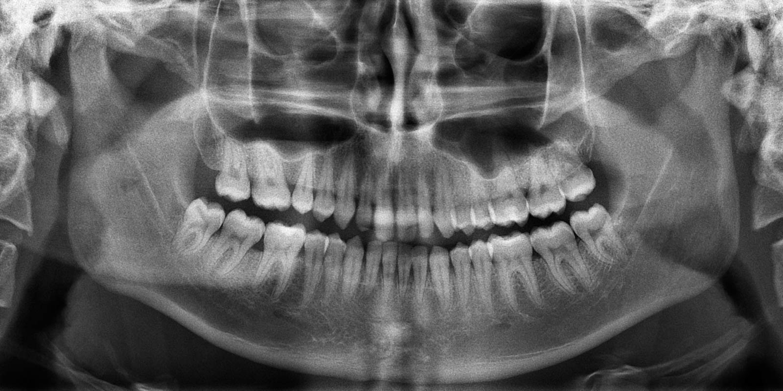
Panoramic images acquired with X-VIEW 2D PAN have a precise focus throughout the entire arch and facilitate the general evaluation of the quantity and quality of bone, dentition and TMJ.
FEATURES
Remarkable features that
cannot be seen:


Available in three models to offer complete and affordable imaging solutions:


X-VIEW 2D PAN UP
This model grows along with the workflow. Initially the doctor acquires an X-VIEW 2D PAN UP that in a second stage becomes a 2D PAN CEPH.
X-VIEW 2D PAN UP, born as a 2D PAN BASIC model with the specific mechanical and user interface dispositions to articulate the cephalometric option in a second stage.
The upgrade process consists of adding the cephalometric arm and a dedicated DR single shot sensor for cephalograms.


GO TO THE NEXT LEVEL WITH FOCUS

Boost your professional practice with the latest technology in digital 2D images acquisition system. The new X-VIEW 2D PAN FOCUS with Image Reconstruction Technology completely exceeds traditional methods to capture panoramic images.
FOCUS technology acquires 2D images derived from the CBCT 3D technology by applying the tomographic approach to OPG field. Newly acquired images are so sharp and clear that they do not need further process.
During the image acquisition, the autofocus function identifies those zones where the interest region is optimally positioned providing images with the best possible sharpness and resolution in no time and less radiation doses.
X-VIEW 2D PAN FOCUS exceeds the most demanding expectations of doctors; the dynamism, ease of use and flexibility of the new technology will always allow them to obtain excellent images even for the most complex cases.

No autofocus

Autofocus

The FOCUS technology is purely based on mathematical operations and the proper calibration of the reconstruction parameters during the X-ray system development phase. The determination of both a proper system and the reconstruction parameters, ensures that a volume of planes spans all the parts of the patient’s relevant anatomy with planes dense enough to ensure that any part of the relevant anatomy is captured with adequate sharpness at least in one of the planes in the reconstructed stack.
-

PROJECTION FRAME
Individual images acquisition from the detector with offset, gain and defect pixel correction. -

TOMOGRAPHIC PLANE RECONSTRUCTION
A set of tomographic planes is created by combining the individual projection frames through a so-called “shift-and-add” method. This process is performed in parallel for several focal planes, using different pixel shift values. -

ANATOMY CONDITIONING
Clinically irrelevant features in select tomographic planes can be suppressed, to reduce their visibility in the final reconstructed image. -

2D IMAGE SYNTHESIS
From the full set of (conditioned) tomographic planes, a single 2D image is synthesized that combines the sharpest information from the total set of tomographic planes throughout different regions of the image. Vertically, regions from planes at different depth positions can be combined to follow complex patient anatomy (e.g. overbite). -

GEOMETRY CORRECTION
The 2D reconstructed image can be scaled in the scan direction, to adjust for magnification and scaling differences due to system geometry and scan motion profiles.
Reviews
There are no reviews yet.